Portable 5 Lead Medical ECG Machine
£312.61 GBP
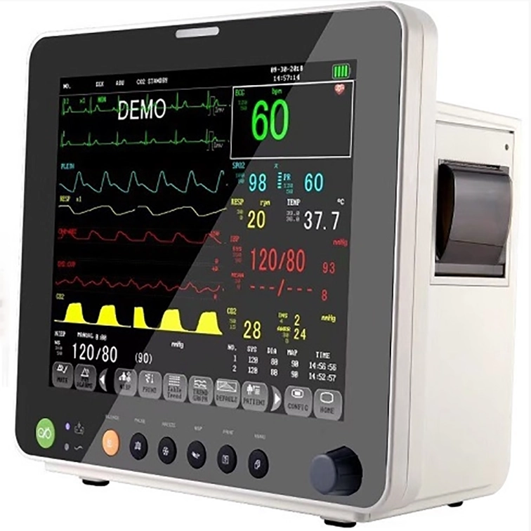
Introducing our medical multi-parameter heart disease monitor with exquisite appearance and multiple functions. This product has the functions of detecting RESP, ECG, TEMP, SpO2, PR, NIBP in adults and children, to help heart disease patients get accurate real-time monitoring of heart condition and fully understand their health status and do a good job in the treatment of heart disease and other related diseases. The machine is supported to be used in hospital and under the guide of doctors.
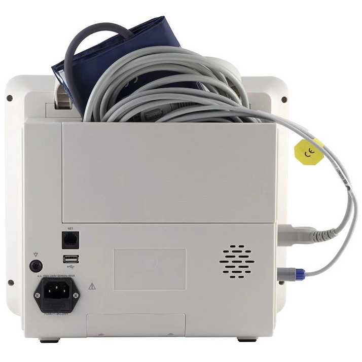
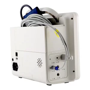
- There is a unique management cabinet in the back of the cardiac monitor host to hold accessories.
- 12.1 inch high resolution color TFT touch screen display, simple and friendly to operate and observe data.
- USB interfaces support easy software upgrade and data transfer.
- Three working modes: monitoring, surgery and diagnosis.
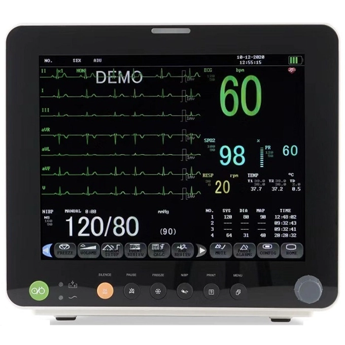
- Multi-display modes suitable for different applications: Standard Interface, Large Font, ECG Standard Full Display.
- Built-in lithium battery for up to 3 hours of continuous work. Both can be plugged in and recharged.
- VGA output for external display, direct to observe data.
How to Connect
Brief Introduction
The detection principle of our 5 lead medical ECG machine is the same as that of electrocardiographs used in hospitals, but they have the advantages of easy portability, simple operation, timely detection, and adaptive adjustment of ECG display amplitude. It provides an effective detection method for detection of heart health situation, and provides doctors with effective information related to patients.
There are three working modes: monitoring, surgery and diagnosis.
- Monitoring: when the patient can lie flat and the monitor has sufficient power or can be plugged in, this product can be used to monitor the patient’s heart condition in real time, thereby upgrade remedy method and make a reference for next treatment.
- Surgery: this monitor is able to be used in the operation of surgery to monitor patient’s heart condition and other physiological indicators, supporting every surgery goes soomth. Diagnosing heart rhythm and check out the success of surgery.
- Diagnosis: this monitor is also can be used to diagnose patient’s kind of heart disease.
Product Details
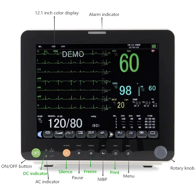
Multiple-functions Display
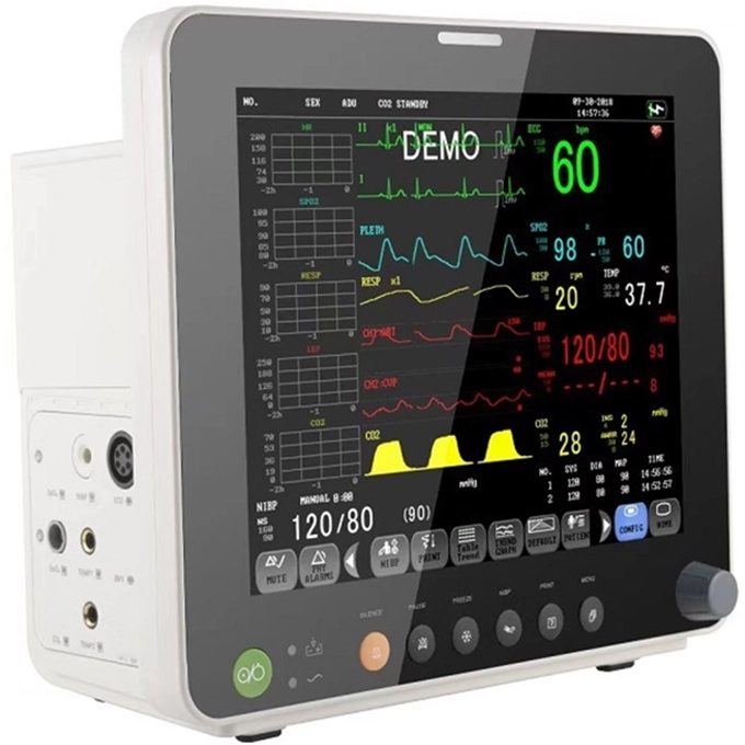
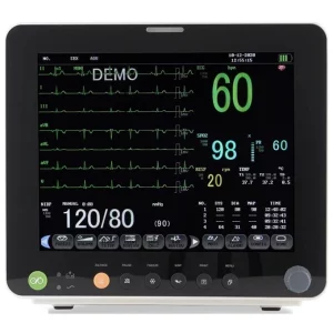
The cardiac monitor can measure a variety of parameters. Most of function buttons are displayed on a touch screen. Each button is marked with a text explanation, which is simple and easy to understand and operate, giving users a comfortable experience.
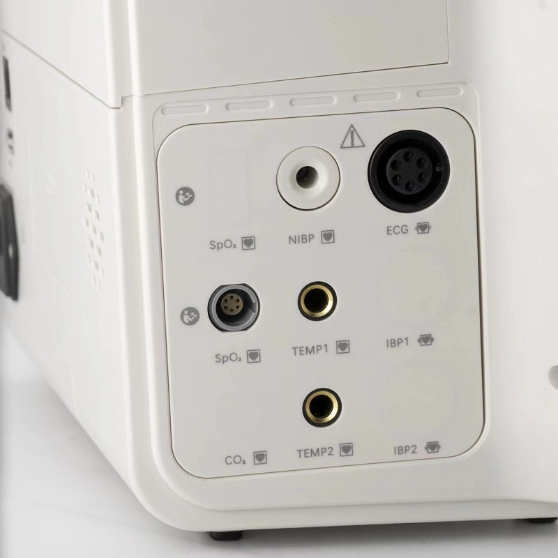
Intuitive Interface Area
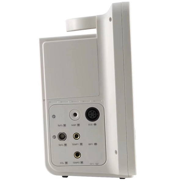
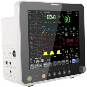
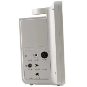
The left side of the monitor host is the interface area. All interfaces are in the same area, which makes it easy for users to quickly find the wire interface and seize the rescue time in an emergency.
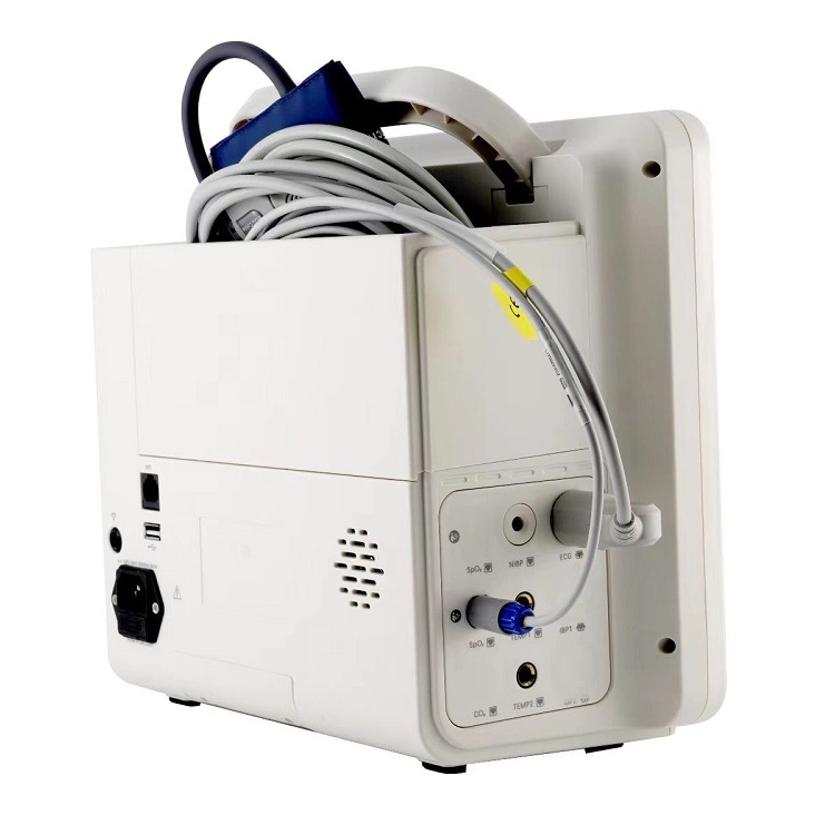
Unique Cabinet for Managing Accessories
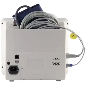
There is a groove on the back of the monitor that can be used as a storage cabinet to store cables, blood pressure cuffs and other accessories, which is convenient and saves space.
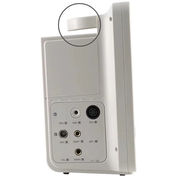
Portable Retractable Handle
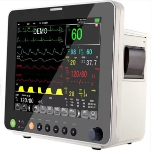
There is a 90° retractable handle on the top of monitor host, convenient to carry it and decreasing the risk of falling and breaking. When needing carry the host, just pull the handle upward 90°, otherwise just fold it back 90°.

Built-in Lithium Battery
Built-in lithium battery supports use after being fully charge, which is convenient for moving patients without cutting off monitoring or treatment. Being fully charged, it can be used lasting for 3 hours. It also cna be plugged in for continuous work.
Product Applications
- Analysis of Patient’s Heart Rate
The ECG machine can help doctors understand the patient’s heart rate more intuitively, thereby making a more accurate analysis and judgment of the patient’s heart rate, which provides a basis for the judgment of arrhythmia.
- Presenting Myocardial Damage and Ventricular Function
Using the ECG machine can more objectively utilize the electrocardiograph curve to reflect the degree of myocardial damage of the patient, and it can also reflect the development process of myocardial damage. The presentation of the electrocardiograph can also reflect whether some adverse changes have occurred in the functional structure of the patient’s ventricles and atria, thereby making a better judgment on the patient’s condition.
- Heart Surgery
During the operation, the ECG machine can monitor the patient’s real-time condition and provide important instructions for the doctor’s operation process to ensure the smooth progress of the operation.
The following groups should pay more attention to their heart health:

People with a family history of heart disease and heart disease
People like this have a higher incidence of heart disease. The incidence rate for men is 2.6 times that of normal people, and for women it is 2.3 times that of normal people. They should be more vigilant and monitor their heart condition regularly.
People with high cholesterol
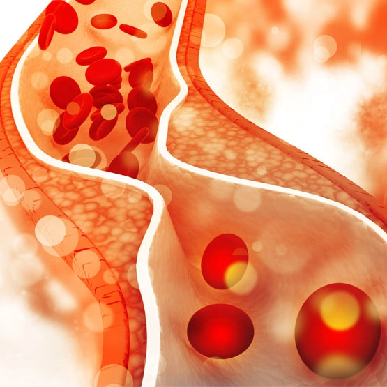
If there is always a large amount of fat and cholesterol in the blood, they will continue to accumulate in the blood vessels, causing the blood vessels to narrow, and the wear rate of the heart will increase exponentially.
People with high blood pressure
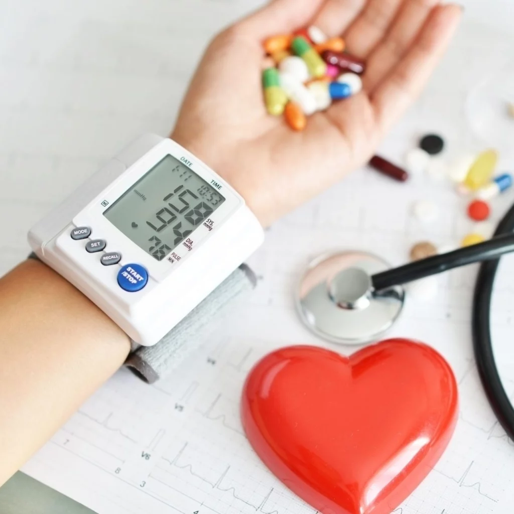
If people don’t take care of their high blood pressure for a long time, it can cause cerebral hemorrhage and sudden death. Compared with normal people, the incidence of heart disease in patients with high blood pressure is about 5-7 times higher.

People with obesity (especially abdominal obesity)
Obese people are more likely to develop high blood pressure, diabetes, dyslipidemia, and insulin resistance syndrome. These adverse effects make their impact on arteriosclerosis not negligible.
People suffering from diabetes
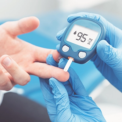
Diabetes can lead to early arteriosclerosis, coronary artery vascular disease, and coronary heart disease. 80% of diabetic patients eventually die of cardiovascular and cerebrovascular diseases. The number of people over 45 years old who die of heart disease is 10-20 times higher than that of non-diabetics.

People like smoking and drinking
The ingredients in cigarettes can damage the heart and blood vessels, causing cracks in the blood vessels and accumulation of fat or cholesterol. Smokers are 2.5 times more likely to suffer from cardiovascular disease than the average person. Long-term heavy drinking can cause myocardial damage, leading to various arrhythmias and symptoms of heart failure.

People with high pressure and mental stress
When the mind is in a state of anxiety for a long time, the body will secrete a large amount of hormones, accelerate breathing and heart rate, and at the same time, the body will release more high-energy fat into the blood vessels, making the blood prone to thrombosis, which will cause myocardial infarction.

People lacking of exercising
People who lack exercise often cannot cope with the physical condition when they encounter an emergency.

Female in the workplace
Women in the workplace have a lot of pressure in life and work. They have to take care of their children, the elderly, and their husbands at home, do housework, and take care of their daily lives. They also have to work hard at work. Compared with men, they put more effort into their careers and families, which makes some women a high-risk group for heart disease.
Specification
ECG (Electrocardiogram)
- Lead Mode: 5 leads (I, II, III, AVR, AVL, AVF, V)
- Gain: 2.5mm/mV, 5.0mm/mV, 10mm/mV, 20mm/mV
- Heart Rate: 15-300 BPM (Adult); 15-350 BPM (Neonatal)
Resolution: 1 BPM
Accuracy: ±1%
Sensitivity: >200uV (Peak to peak) - Sweep Speed: 12.5 mm/s, 25 mm/s,50mm/s
- Bandwidth:
Diagnostic: 0.05-130 Hz
Monitor: 0.5-40 Hz
Surgery: 1-20 Hz
NIBP (Non-invasive Blood Pressure)
- Method: Oscillometry
- Measure Mode: Manual, Auto, STAT
Measuring Interval in Auto Mode: 1, 2, 3, 4, 5, 10, 15, 30, 60, 90, 120, 180, 240,480 (Min)
Measuring Period in STAT Mode:5 Min - Pulse Rate Range:40 ~ 240 bpm
- Alarm Type:SYS, DIA, MEAN
- Measure and Alarm Range:
Adult Mode:
SYS: 40 ~ 270 mmHg
DIA : 10 ~ 215 mmHg
MEAN: 20 ~ 235 mmHg
Pediatric Mode:
SYS: 40 ~ 200 mmHg
DIA : 10 ~150mmHg
MEAN: 20 ~ 165 mmHg
Neonatal Mode:
SYS: 40 ~ 135 mmHg
DIA : 10 ~100mmHg
MEAN: 20 ~ 110 mmHg - Overpressure Protection
Adult Mode: 297±3 mmHg
Pediatric Mode: 240±3 mmHg
Neonatal Mode: 147±3 mmHg
SpO2 (Blood Oxygen Saturation Detection)
- Measuring Range:0 ~ 100 %
- Alarm Range:0 ~ 100 %
- Resolution:1 %
- Accuracy:
70% ~ 100% ±2 %
0% ~ 69% unspecified - Actualization interval: about 1 Sec
- Alarm Delay: 10 Sec
RESP (Respiratory Rate)
- Method: Impedance between R-F(RA-LL)
- Differential Input Impedance: >2.5 MΩ
- Measuring Impedance Range: 0.3~5.0 Ω
- Base line Impedance Range: 0 – 2.5 KΩ
- Bandwidth: 0.3 ~ 2.5 Hz
- Measuring and Alarm Range
Adult: 0 ~ 120 rpm
Neo/Ped: 0 ~ 150 rpm
Resolution: 1 rpm
Accuracy: ± 2 rpm
Apnea Alarm: 10 ~ 40 S
TEMP (Body Temperature)
- TEMP Measuring and Alarm Range:0 ~ 50 °C
- Resolution: 0.1°C
- Accuracy: ± 0.1°C
- Actualization interval: about 1 Sec
- Average Time Constant: < 10 Sec
PR (Pulse Rate)
- Measuring and Alarm Range: 0~254 BPM
- Resolution: 1 BPM
- Accuracy: ± 2 BPM
Notice
- There are no components which can be repaired by customers. Please do not disassemble them when failure occurs.
- This monitor is not a common therapeutic device, please use it as per professional guide.
- Defibrillation is not accessible to patients, beds and this monitor.
- Before cleaning monitor, the power supply of the net must be cut off.
- The monitor shall not be used in places with high temperature, high humidity, inflammable, excessive dust and electromagnetic radiation.
- Ensure the safety and stability of the power grid and grounding environment of this instrument.
- Other details are provided in the manual.
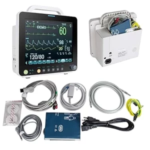
Packing List
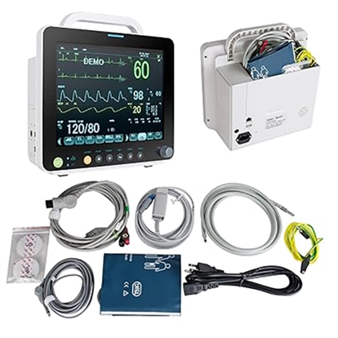
- 1 × Monitor
- 1 × ECG Lead wire
- 1 × NIBP Cuff
- 1 × NIBP Extension Cable
- 1 × SpO2 Sensor
- 1 × Temp Sensor
- 1 × Ground Cable
- 5 × Disposable ECG Electrode
- 2 × Fuse
- 1 × User Manual
NOTE: All default accessories are for adults, please make a note when you place an order if accessories you need are for babies.
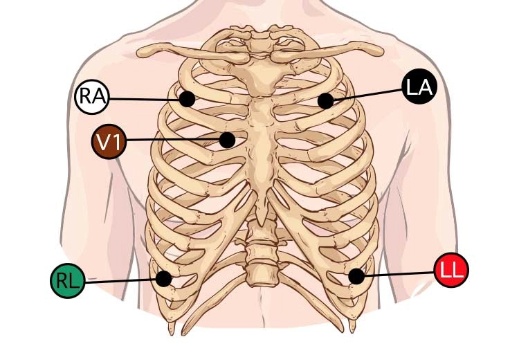
ECG electrodes are colour-coded, and each is identified by a specific code that refers to its intended placement. There are two coding systems currently in use:
- The American Heart Association (AHA) system
- The International Electrotechnical Commission (IEC) system.
Note: The following guide uses the AHA system.
1. Prepare items
Mainly include ECG monitor, ECG and blood pressure plug-in connecting wires, 5 electrodes, saline cotton balls, and matching blood pressure cuffs.
2. The operating procedures are as follows
(1) Connect the power supply of the ECG monitor.
(2) Place the patient in a supine or semi-recumbent position.
(3) Turn on the main switch.
(4) Wipe the patient’s chest skin where the electrodes are attached with 75% ethanol cotton balls.
(5) Attach the electrodes, connect the ECG lead wires, and the ECG waveform will appear on the screen.
(6) Select a suitable cuff and tie the cuff to two horizontal fingers above the elbow.
(7) Set the alarm limit and measurement time.
| RA placement | WHITE directly below the clavicle and near the right shoulder. |
| LA placement | BLACK directly below the clavicle and near the left shoulder. |
| RL placement | GREEN on the right lower abdomen. |
| LL placement | RED on the left lower abdomen. |
| V placement | BROWN on the chest – the position depends on your required lead selection (4th intercostal space, the right side of the sternum). |
- Connect lead wires to the ECG connector and ensure the waveform is visible on the monitor.
- Ensure all monitor alarms are on and audible.
- Always check with a professional if unsure.
- Replace the electrodes at least daily, or more frequently as required.
Only logged in customers who have purchased this product may leave a review.

Reviews
There are no reviews yet.